High-Content Analysis Systems
A selection of powerful High-Content Analysis Systems combined with intuitive user software to provide flexible research applications.
Yokogawa’s High Content Analysis Systems (HCA), also known as High Content Screening Systems (HCS), lead the microscopy industry in higher product quality, personalized process control, and streamlined user interfaces. With over 25 years of experience and a track record of proven success, we are ready to help you exceed your goals.
-
CellVoyager High-Content Analysis System CQ3000
CQ3000 can acquire high-resolution 3D images at high speed while culturing cells by combining options according to the application.
Details
What is High Content Analysis (HCA)?
High-content analysis is a principle methodology for so-called phenotypic screening in drug discovery and development process. Assessing a vast number of compounds is time consuming, therefore, acquiring a large number of high resolution cellular images with fast scanning speed is critically important for drug screening. Yokogawa high-content analysis imagers have balanced these generally conflicting requirements at a high level of performance. The core technology, the microlens-enhanced dual Nipkow disk in Yokogawa Confocal Scanner or Confocal Spinning disc Unit (CSU), makes it possible to capture high quality images with remarkably short acquisition time.
In addition to the image acquisition unit, a precisely controlled fast moving automated stage chamber is also an essential component of our imaging systems. Monolayer fixed cell samples have been commonly used in drug screening. However, cellular environments in such fixed 2D samples are quite different from those of actual live systems, and the divergence often causes a significant discrepancy between experimental results and what is observed in real organisms.
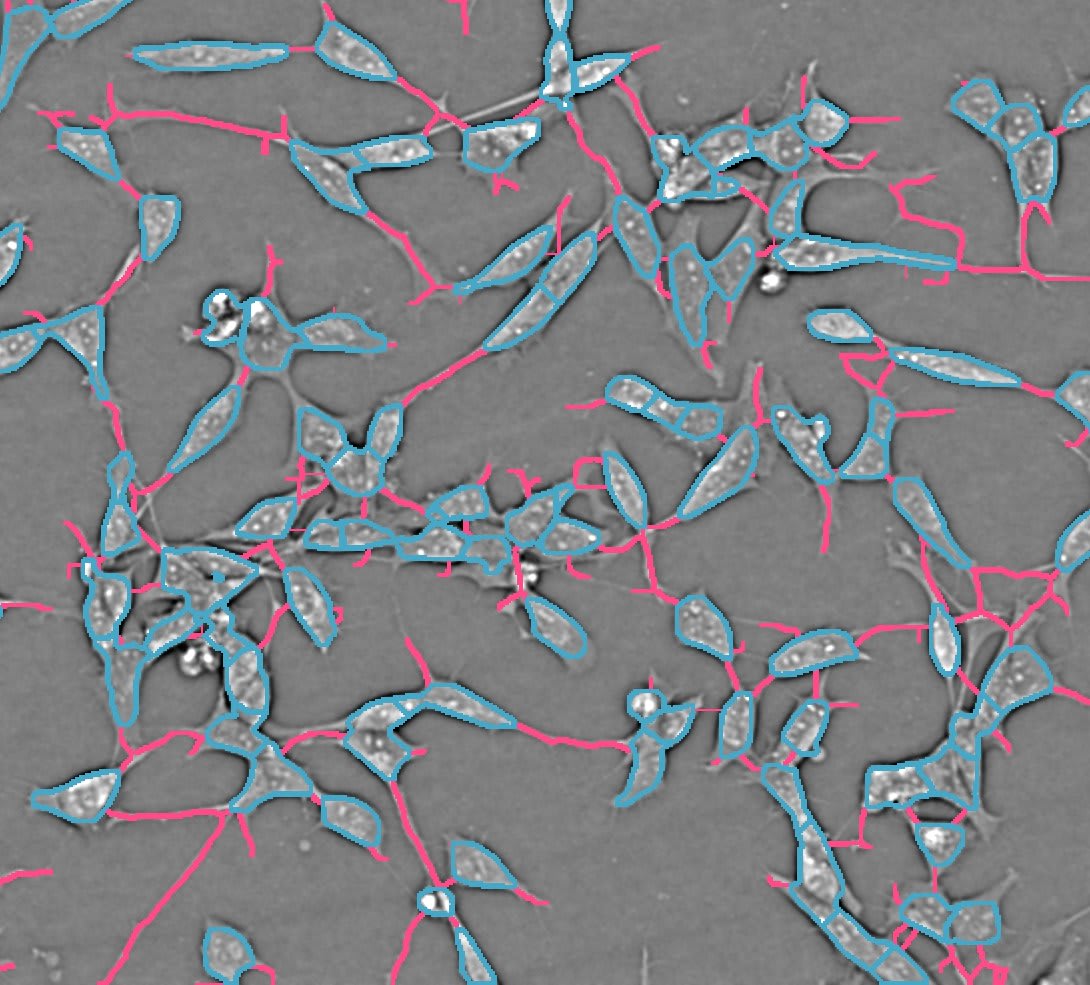
These days, using 3D samples such as spheroids and organoids, and live cell samples to mimic realistic cellular environments are becoming more and more popular due to cell culture technology development. Yokogawa CSU is renowned in the microscopy field not only for the high image quality and fast capturing speed, but also for the live cell friendliness. Our high-content confocal imaging systems based on the CSU are the ideal solution for drug screening using elaborated 3D and live cell assay systems as well as 2D fixed samples. User friendly high content analysis software CellPathfinder, with its versatile functionalities, powerfully supports the analysis of various phenotype changes and target reactions. It also has state-of-the-art machine learning and deep learning function for complex sample analysis. All the technologies are packed into the Yokogawa high-content analysis system.
High Content Analysis vs Traditional Methods
| Traditional Methods | High Content Analysis (HCA) | ||
|---|---|---|---|
| High-Throughput Screening (HTS) | Flow Cytometry (FCM) | Microscopy | |
|
Strength:
Weakness:
|
Strength:
Weakness:
|
Strength:
Weakness:
|
Strengths:
|
Confocal Imagers with Powerful Analysis Software
Our high content analysis (HCA) systems are the best solutions for a range of research applications from basic science to drug discovery screening.
Resources
PhenoVista Biosciences is the leading provider of custom, imaging-based, phenotypic assay services. With a collaborative and scientifically driven project design and management approach, PhenoVista has a proven track record of delivering high-quality data from robust and scalable assays. PhenoVista’s key advantage lies in the ability of their industry-trained scientists to combine world-class understanding of diverse biological systems with cutting-edge quantitative imaging to deliver clear, actionable output data.


Cell stage categorized using FucciTime lapse imaging of Fucci-added Hela cells was conducted over 48 hrs at 1 hr intervals. Gating was performed based on the mean intensities of 488 nm and 561 nm for each cell. They were categorized into four stages, and the cell count for each was calculated.


The CV8000 nuclear translocation analysis software enables the analysis of changes in the localization of signal molecules that transfer between cytoplasm and nuclei, such as proteins. The following is an example of the translocation analysis of NFκB, a transcription factor.


The CQ1 confocal image acquisition mechanism with the distinctive CSU® unit has a function to sequentially acquire fine cell images along the Z-axis and capture information from the entire thickness of
cells which include heterogenic populations of various cell cycle stages. In addition, saved digital images can be useful for precise observation and analysis of spatial distribution of intracellular molecules.
The CQ1 capability to seamlessly analyze images and obtain data for things such as cell population statistics to individual cell morphology will provide benefits for both basic research and drug discovery
targetingM-cell cycle phase.


In this tutorial, we will learn how to perform cell tracking with CellPathfinder through the analysis of test images.

In this tutorial, using images of zebrafish whose blood vessels are labeled with EGFP, tiling of the images and recognition of blood vessels within an arbitrary region will be explained.
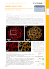
In this tutorial, a method for analyzing ramified structure, using CellPathfinder, for the analysis of the vascular endothelial cell angiogenesis function will be explained.

In this tutorial, we will learn how to perform time-lapse analysis of objects with little movement using CellPathfinder, through calcium imaging of iPS cell-derived cardiomyocytes.

In this tutorial, we will observe the change in number and length of neurites due to nerve growth factor (NGF) stimulation in PC12 cells.

In this tutorial, image analysis of collapsing stress fibers will be performed, and concentration-dependence curves will be drawn for quantitative evaluation.
In this tutorial, we will identify the cell cycles G1-phase, G2/M-phase, etc. using the intranuclear DNA content.

In this tutorial, intranuclear and intracytoplasmic NFκB will be measured and their ratios calculated, and a dose-response curve will be created.

In this tutorial, spheroid diameter and cell (nuclei) count within the spheroid will be analyzed.

In this tutorial, a method for analyzing ramified structure, using CellPathfinder, for the analysis of the vascular endothelial cell angiogenesis function will be explained.
Downloads
Videos

The CV8000 features a cell incubator with an improved airtight design that facilitates the observation of cell behavior over long periods of time. In addition, the CV8000 comes with CellPathfinder, a new program that can analyze images of unlabeled cells and 3D images of samples. With these features, the CV8000 improves the efficiency of drug discovery research and biomedical research on leading-edge subjects such as iPS and ES cells.

Watch the film by KBC who are supporting their clients in their efforts to address climate change and sustainability.
Looking for more information on our people, technology and solutions?
Contact Us