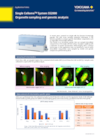
SU10 and SS2000 are groundbreaking systems designed for precise single-cell manipulation
These innovations enable the introduction of substances into cells, allow for observation before and after, and facilitate the extraction of intracellular components. With their flexibility and versatility, these accelerate research across diverse fields, including biological phenomena, disease mechanisms, drug discovery, and the development of new plant varieties. Additionally, our dedicated support team is always available to address challenges and drive success.
-
Nano-point Delivery/Nano-point Sampling Single Cellome Unit™ SU10
Single Cellome Unit SU10 enables the delivery of substances, such as recombinant proteins and genome editing tools, directly to the cytoplasm or nucleus of targeted single cells.
-
Single-cell and Subcellular Sampling System
The SS2000 is a dual microlens spinning disk confocal microscope for live-cell, high-content imaging which can sample adherent single cells and subcellular material while ensuring the preservation of spatial, morphological, and temporal information.
Details
Nano-point Delivery / Nano-point Sampling
Single Cellome Unit SU10 enables selective delivery into the nucleus and cytoplasm of targeted cells under a microscope.
Using a glass pipette (nanopipette) tip with a diameter of several tens of nanometers minimizes damage to cells.
Single Cellome Unit SU10 can deliver various substances or sample from specific parts of cells at high speed and efficiency by automating cell surface detection, puncture, as well as injection and sampling operations.

Subcellular Sampling
The Single Cellome System SS2000 can automatically sample intracellular components and whole cells under a confocal microscope.
Since cells in culture do not need to be detached, it is possible to sample while maintaining positional and morphological information.
This sampling system combines high content analysis functions such as high-speed, high-resolution image acquisition and imaging analysis.

Resources
The SU10 is a novel technology that can deliver target substances into cells (nucleus or cytoplasm) by using a "nano" pipette made of glass capillary with an outer tip diameter of just tens of nanometers.
The SU10 is a “nano” pipette with a tip outer diameter of only a few tens of nanometers, which can automatically deliver target substances into cells (nucleus, cytoplasm) at the single-cell level. The SU10 can directly deliver genome editing tools into the nucleus, so it is expected to enable efficient genome editing. In this application note, we will introduce a case study in which Cas9 RNP and donor DNA were delivered into cells using SU10, resulting in the successful knockout (KO) and knock-in (KI) of target genes.
The SS2000 is a revolutionary system that allows both confocal imaging and sampling of targeted intracellular components or single cells using a glass tip with an inner diameter of a few micrometers. The SS2000 includes an incubator as well as highcontent imaging functions, such as long time-lapse observation, machine learning, and label-free analysis.
The SU10 is an innovative device that delivers target substances directly into cells or into nuclei with a nanopipette that has a tip outer diameter as small as a few tens nm.
This application note shows a case study of sampling and analysis with the SS2000 from cells delivered intracellularly with the SU10.
In recent years, single-cell studies have become increasingly popular, leading to the development of instruments capable of spatial omics - the integration of samples' 3D locations and their omics data. The SS2000 is a revolutionary device that can collect intracellular components at the single-cell level and image the samples using a confocal microscope. In this application note we introduce the research performed by Dr. Okada of The University of Tokyo, who established an intra single living cell sequencing (iSC-seq) method which uses SS2000 to sample intracellular components from osteoclasts (a type of multinucleated giant cell) and analyzes them through next-generation sequencing.
In recent years, research on single cells has been actively conducted.However, with more sensitive analytical techniques, it is now possible to analyze specific intracellular components at the single-cell level.
In recent years, research on single cells has become increasingly popular, but with more sensitive analytical techniques, it has become possible to analyze specific intracellular components such as organelles at the single-cell level.
The SS2000 is an innovative system that can sample intracellular components at the single-cell level using a tip (glass capillary) with a diameter of several micrometers while imaging with a confocal microscope. In this application note, we introduce a case in which intracellular components were sampled by SS2000 and genetic analysis was performed by qPCR.
SU10 is a novel technology that enables the delivery of target substances directly into cells (nucleus or cytoplasm) using a "nano" pipette made of a glass capillary with an outer tip diameter of tens of nanometers.
In recent years, single cell analysis has become increasingly popular due to the new development of advanced and high sensitivity analytical methods.
SS2000 is a revolutionary system that can sample subcellular components and a whole cell by using a glass capillary tip with an inner diameter of a few μm while imaging with a confocal microscope.
This application note provides an example of single cell RNA sequencing scRNA seq) from cells sampled by SS2000, and the data is comparable to conventional methods.
In recent years, research on single cells has become increasingly popular. With more sensitive analytical techniques, it has become possible to analyze specific intracellular components such as organelles, even at the single-cell level.
The SS2000 is an innovative system that can sample subcellular intracellular components using a glass capillary having a tip diameter of only a few micrometers while imaging with a confocal microscope.
In this application note, we introduce a case where after drug treatment, intracellular components were sampled by the SS2000 and analyzed by single-cell mass spectrometry.
Videos
This webinar highlights Yokogawa’s High Content Solutions, the benchtop confocal CellVoyager CQ1, and CellVoyager CV8000. Utilizing Yokogawa’s dual-wide microlens spinning disk confocal technology, these automated HCA systems provide remarkable image quality while increasing your output. This frees up time to complete other research activities. Also, recent additions to the CSU-W1 confocal upgrade is discussed. The SoRa, a super-resolution solution, and the Uniformizer, an image flattening device. Both of which can be added to the lightpath of your CSU-W1-enhanced microscope.
Agenda:
Introduction to Yokogawa
SoRa for CSU-W1 super-resolution with confocal
Two high content instruments from Yokogawa: The CQ1 and the CV8000
Presenter:
Dan J. Collins, Applications Scientist, Yokogawa Life Science
In the last few decades, the pharmaceutical industry has transformed people’s lives. However, the development of new drugs is becoming increasingly difficult and a paradigm shift in the drug discovery workflow is required to reduce attrition and transform conventional drug screening assays into translatable analytical techniques for the analysis of drugs in complex environments, both in-vitro and ex-vivo. The ability to visualize unlabelled compounds inside the cell at physiological dosages can offer valuable insight into the compound behavior both on and off-target.
SiLC-MS is a semi-automated methodology that allows the collection of intracellular contents using a modified CQ1 imaging system developed by Yokowaga. The instrument is equipped with a confocal microscope that allows bright field imaging as well as fluorescence imaging with 4 lasers (405, 488, 561, and 640 nm). Sampling is performed using the tips developed by Professor Masujima (1-4).
In this study, we show the applicability of the SiLC-MS technology to drug discovery, as it is crucial to identify compound and its metabolites when incubated in a mammalian cell at a therapeutic dose. We report on the validation studies performed using the SiLC-MS platform, in these validation studies we assess the ability to distinguish different cell types based on their metabolomic fingerprint, furthermore, we have also evaluated if this assay was sensitive enough to detect drugs intracellularly.
Presenter: Carla Newman, Scientific Leader (Celluar Imaging and Dynamics), GSK
News
-
News Brief Apr 25, 2024 Yokogawa Helps to Revolutionize the Field of Single-Cell Lipidomics
-
Press Release | Solutions & Products Nov 30, 2021 Yokogawa Develops Single Cellome System SS2000 for Subcellular Sampling
Looking for more information on our people, technology and solutions?
Contact Us


