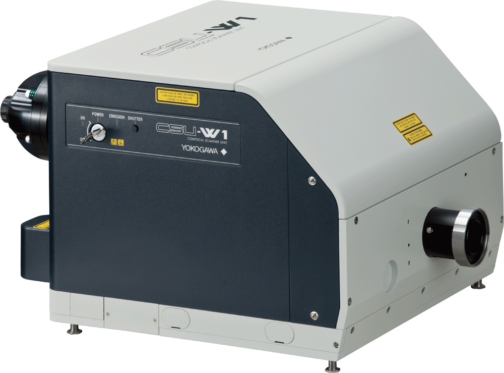
Widest field of view in the industry providing 4X wider FOV than conventional scanners.
The CSU-W1 Confocal Scanner Unit is our widest field of view confocal, providing the clearest image resolution of our imaging systems. The system features switching mechanisms to enable fully automated experiments and a newly designed disk unit to improve image clarity of thick samples.
New option CSU-W1 Uniformizer is released. About Uniformizer
- Wide and clear
- Near Infrared (NIR) Port: Up to 785nm
- 3 configurations: 1-camera model, 2-camera model, split-view model
- New bright field path (Standard)
- Selectable pinhole size: 25 pinhole disk, 50 pinhole disk or double disk
- External light path
- 10-position filter wheel (1 Camera model, 2 Camera model)
- Fully automate experiments with the motorized switching mechanism
Wide
Widest FOV confocal! Provides 4 times wider FOV than the conventional model.

Clear
Newly designed disk unit offers much improved image quality. Due to significantly reduced pinhole crosstalk, CSU-W1 enables clear observation much deeper into thick samples.
Conventional model
|
XY MIP |
XZ Slice |
 |
 |
CSU-W1
|
XY MIP |
XZ Slice |
 |
 |
Mouse ES cell colony
Fluorescent probe: H2B-EGFP(Excitation:488nm) mCherry-MBD-NLS(Excitation: 561nm)
Objective lens: 60x silicone
Z-sections/stack: 100um(0.4m/251slices)
By courtesy of Jun Ueda, Ph.D. and Kazuo Yamagata, Ph.D., Center for Genetic Analysis of Biological Responses, The Research Institute for Microbial Diseases, Osaka University

Flexible
Flexibly selectable functions to meet versatile applications.
High confocality pinhole (Optional Component)
In addition to our conventional 50um pinhole size, 25m pinhole size with higher confocality is available.
You can select either one or the both pinhole size, with easy-to-use motorized disk exchange mechanism.

New bright field through path (Standard)
New mechanism to move the disks out of the light path allows much easier projection of confocal and non-confocal images such as phase contrast.
Simultaneous dual color imaging mechanisms
(T2 and T3 Models)
CSU-W1 offers single camera split-view model, in addition to the dual camera model which are much improved from those for the CSU-X1. Thanks to the wide FOV, even the split-view offers 2 times wider image area than with older model. By using various dichroic mirrors, it is possible to select various dye-combinations for dual-color imaging*1 with both the two camera model and split-view model.
Details
CSU-W1 offers selection from a total of three basic configurations, two pinhole sizes, options for near infrared observation and an external light path which is useful for versatile applications such as photo bleaching, while bright field light path is now a standard feature. All switching mechanisms in the CSU-W1 are fully motorized and thus ready for automated experiments.
Basic Configurations
CSU-W1 provides a total of three basic configurations for multi- color imaging; 1) Sequential imaging with one camera and a filter wheel, 2) Simultaneous two-color imaging with two cameras, and 3) Split-view two color imaging with one camera shared by 2 optical paths. All features are upgradable after installation.

Filter

Option
SoRa disk
Optical resolution has been improved by approximately 1.4x using a super-resolution technique based on spinning-disk confocal technology. Furthermore, a final resolution approximately twice that of the optical limit is realized through deconvolution.
Upgrading from the CSU-W1:CSU-W1 SoRa
Uniformizer
Most suitable option for CSU-W1 illumination uniformity. Uniform and efficient illumination of the entire wide field of view.
About Uniformizer:More info
Near Infrared (NIR) Port
NIR port provides up to 785nm excitation capability to allow less-invasive deep imaging. The NIR laser is introduced via a dedicated optical fiber in the same way as visible lasers. It is possible to combine NIR and visible lasers within the CSU-W1 unit to allow simultaneous excitation.
External light path
External light path provides the direct path bypassing the disk s to microscope. Versatile applications such as photo activation are available by introducing an external light scanner through this port.
Lens switcher
Newly designed motorized lens switcher between 2 relay lenses i s useful for fitting CSU-W1 image size with various camera types, and also for easy magnification change without exchanging objective lenses.
Variable aperture
Variable aperture to change laser illumination area, and thus the imaging area by the CSU-W1, is useful to minimize laser damages in the specimen.
Selectable option
| Option | 1 Camera model | 2 Camera model | Split-view model |
|---|---|---|---|
| NIR port | × | ||
| External light path | × | ||
| Variable aperture | × | N/A | |
| Camera port lens | Selectable from 0.83x, 1x |
Selectable from 0.83x, 1x (1 Camera) Selectable from 0.83x, 1x (2 Camera) |
Selectable from 0.83x, 1x |
| Additional lens to lens switcher |
Selectable from 0.83x, 1x, 2x | N/A | |
2 Camera model, 1 Camera model

Split-view model

*1 2 Camera model *2 2 Camera model and Split-view model
*3 1 Camera model and 2 Camera model *4 Under development
External Dimensions

Microscope-setup
|
|
|
|
|
|
|
|
| General Specifications | ||||
|---|---|---|---|---|
| Model | 1 camera model (T1) | 2 camera model (T2) | Split-view model(T3) | |
| Confocal scanning method | Microlens-enhanced Nipkow disk scanning | |||
| Spinning speed | 1,500rpm - 4,000rpm Max200fps | |||
| External synchronization | Scan-speed synchronization through pulse signals Input/output : TTL level 300Hz up to 800Hz | |||
| Disk unit | Selectable up to 2 disks from 50um (for high magnification) and 25um(for low magnification) Motorized switching |
|||
| Bright field | Motorized exchange between confocal and brightfield | |||
| Effective FOV | 17×16mm | |||
| Excitation wavelength | 405nm-785nm | |||
| Laser introduction | Yokogawa's standard fiber*1 VIS Laser port (405-647nm) 【Option】NIR Laser port (685-785nm) |
|||
| Observation wavelength | 420nm-850nm | |||
| Dichroic mirror switching | Motorized 3CH (Dichroic mirror block can be exchanged) | |||
| Emission filter wheel | 10-position filter wheel Filter sizeφ25mm Switching speed*2 : 100msec max.(Standard mode) 40msec max.(High speed mode) |
6-position filter wheel Filter sizeφ25mm Switching speed*2 : 100msec max. |
||
| External control | RS-232C (CSU-X1 command upper compatible) | |||
| Microscope mount | Yokogawa original | |||
| Camera adaptor | C mount 1x (Variable magnification: 0.83x ) | |||
| Light introduction port | 【Option】Photo breach etc | |||
| Operating environment | 15-30oC、20-75%RH No condensation | |||
| Power consumption | Input : 100-240 VAC ±10% 50-60Hz 250VAmax | |||
| External dimension | Main unit | 327.1(W)x 251.5(D)x 475.1(H)mm |
471.6(W)x 251.5(D)x 475.1(H)mm |
420.8(W)x 251.5(D)x 373.6(H)mm |
| Power unit | 225.4(W)x 151.9 (D)x 378.3(H)mm | |||
| Weight | Main unit | 17kg | 20.5kg | 18kg |
| Power unit | 4.5kg | |||
| Attachable microscope | Olympus IX series, Nikon ECLIPSE Ti , Zeiss Axio Observer, Leica DMI series*3 | |||
*1 Each CSU-W1 head is optimized with its fiber at factory. Please inquire about fiber exchange if necessary.
*2 Adjacent position.
*3 Some microscopes/options could limit the FOV of CSU-W1 or connection with CSU-W1, please inquire.
We post our information to the following SNSs.
Please follow us.
Yokogawa Life Science
| @Yokogawa_LS | |
| Yokogawa Life Science | |
| Yokogawa Life Science | |
| •YouTube | Life Science Yokogawa |
Yokogawa's Official Social Media Account List

Nikon Instruments Inc.
(North and South America)
Nikon Instruments Europe B.V.
(Europe)
Nikon Corporation
(Asia except for Japan)
Resources
Wide and Clear
Confocal Scanner Unit
Long-term observation of mitosis by live-cell microscopy is required for uncovering the role of Cohesin on compartmentalized nuclear architecture which is linked to nuclear functions.
To perform long term observation of mitosis devices are needed that have low phototoxic effects on living cells and enable high speed imaging. By using the CSU W-1 confocal scanner unit for time lapse imaging entrance into mitosis, mitotic progression and exit can be examined.
SU10 is a novel technology that enables the delivery of target substances directly into cells (nucleus or cytoplasm) using a "nano" pipette made of a glass capillary with an outer tip diameter of tens of nanometers.
To investigate interactive dynamics of the intracellular structures and organelles in the stomatal movement through live imaging technique, a CSU system was used to capture 3-dimensional images (XYZN) and time-laps images (XYT) of guard cells.
List of Selected Publications : CSU-W1
Downloads
Brochures
- Confocal scanner unit CSU-W1 (2.6 MB)
Videos
YOKOGAWA proprietary Spinning Disk technology enables fast real-time confocal imaging for applications such as high-speed 3D and long-term live cell imaging. These quantifiable imaging analysis are essential tools for modern precision drug discovery.
YOKOGAWA will contribute to technology evolution particularly in measurement and analytical tools to help build a world where researchers will increasingly focus on insightful interpretation of data, and advancing Life Science to benefit humanity.
Over past 20 years, YOKOGAWA proprietary Spinning Disk Confocal technology has been widely used as an indispensable imaging tool among top researchers. The technology enables faster live-cell observation with clearer and less photo-bleaching imaging.
News
-
Press Release Dec 3, 2020 Yokogawa and InSphero Enter into Partnership Agreement
- Supporting drug development research through the use of HCA and three-dimensional culture models -
Looking for more information on our people, technology and solutions?
Contact Us


