Revealing the pathophysiology of COVID-19 infection
For months, the COVID-19 pandemic holds many countries and societies of our globe hostage. Besides attempts to reduce the number of new infections to prevent a collapse of health care infrastructure, the whole world is desperately striving to learn as much as possible about the disease and its causing virus SARS-CoV-2. Many researchers and institutions are performing research trying to find treatment methods for better and faster recovery avoiding lethal or fatal progression. Also, the race for a COVID-19 vaccine is on, moving forward at unprecedented speed.
A bigger footprint
The Life Innovation Business (LIB) and product portfolio are of growing strategic importance for Yokogawa. We are developing and acquiring technologies to leave a bigger footprint in this area. Human well-being is also one of our company’s core values. Yokogawa decided to support the world’s fight against COVID-19, scientifically cooperating with research facilities actively involved in COVID-19 research. In those global collaborations, researchers will use the Yokogawa bench-top confocal quantitative image cytometer – the CellVoyager CQ1 microscope. This instrument is of compact size fitting into every laboratory, it can be set up as well as tested in a day and is easily learned to operate by new users. Our collaborating research groups are distributed over the whole world and we are very happy to have one partner in Berlin, Germany.
COVID-19 research at Charité in Berlin
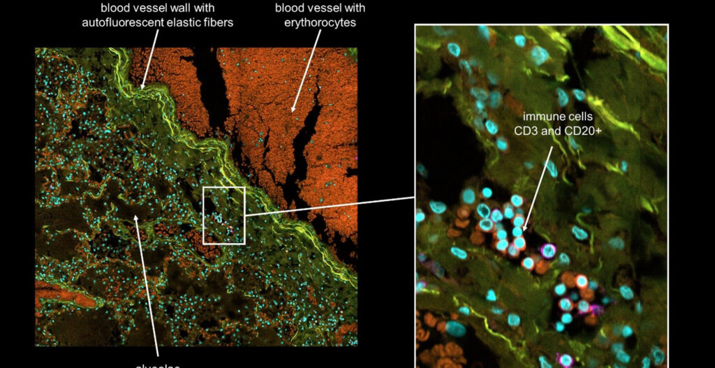
The Charité University Medicine in Berlin is one of the largest hospitals and academic medical centers in Europe playing an important role in the response to the COVID-19 pandemic in Germany. Several ongoing projects at the Institute of Pathology of the Charité University Hospital focus on characterizing the pathophysiology of infections with COVID-19 (Coronavirus SARS-CoV-2) in tissues from COVID-19 infected patients. The projects involve extensive characterization of all affected organ systems. They are focusing on both the dynamics of the inflammatory response to infections with COVID-19 as well as the composition of the immune infiltrate and the tissue damage resulting from this disease using immunohistochemistry.
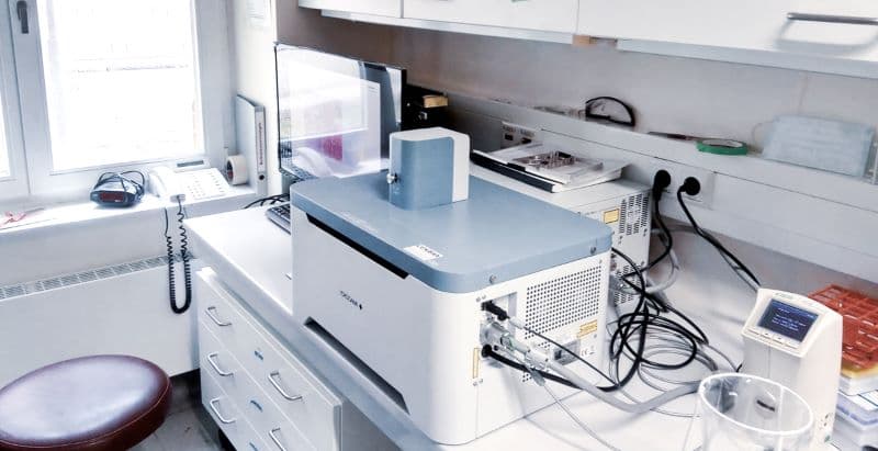
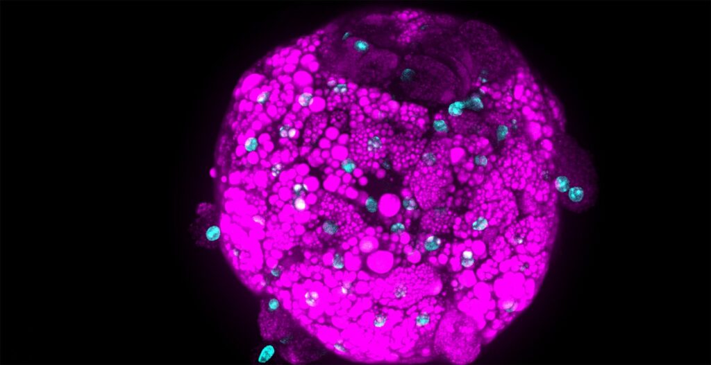
The CellVoyager CQ1 microscope, which can be used for High-Content Imaging, gives the Charité researchers a new possibility to perform multiplexed immunofluorescence multicolor confocal imaging on the same slide significantly improving the throughput and the quality of obtained images compared to conventional widefield slide scanners.
Better image quality
In addition to higher throughput and better image quality that can be achieved using the CQ1, the investigators are looking forward to applying the wide-ranging image analysis capabilities provided by the CQ1 and CellPathfinder™ software suite to analyze the resulting images in a systematic way. This will enable a better understanding of the complex multidimensional datasets generated during the experiments. In summary, the research group around Prof. Horst believes that the opportunity to use the CellVoyager CQ1 microscope provided by Yokogawa in this collaboration will allow a significant acceleration of the COVID-19 related projects, generating data of higher quality, thus contributing to an urgently needed better understanding of the pathophysiology of this disease.
Supporting this research project in Germany and worldwide Yokogawa hopes to contribute to a better understanding of the COVID19 infection. Our High-Content Imaging microscope, analysis software suite and other products can help to move a little step further on the long and slippery road to overcome this global health crisis.
