Introduction
Real-time, simultaneous observation of individual neuronal activity over a large area of the brain is required to understand how the brain perceives external sensory information. To achieve this, devices capable of wide-field imaging with high spatial and temporal resolution are necessary.
The combined use of our CSU-WI confocal scanner and the GCaMP calcium indicator enables imaging of brain activity in zebrafish larvae at a remarkably high resolution.
※Reorganized using the maximum intensity projection (MIP) function.
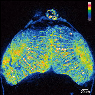
Fig. 1: Real-time observation of neuronal activity in the optic tectum of zebrafish larva *1

Fig1(a).Zebrafish larva five days after fertilization (□: optic tectum)
All time lapse movie: Play

Fig1(b).3D imaging of the brain (tectum) expressing GCaMP

Fig1(c). A GCaMP fluorescence image, single frame extracted from a time-lapse movie.

Fig1(d).Superimposed time-lapse images. Areas with strong calcium reaction are shown in red
Experiments
A Zebrafish larva (three to five days after fertilization) expressing the calcium indicator GCaMP in the optic tectum was fixed in agarose, and imaged the neuronal activity in its tectum of both the spontaneous one, and of the response to a visual stimuli.

| Sample | UAS (Gal4-UAS system*2): GCaMP7a zebrafish larva three to five days after fertilization |
| System | Confocal unit: CSU-W1 (pinhole: 50μm) Laser: 488nm (solid-state laser) Microscope: Axio Imager (Carl Zeiss) Camera: iXon 888 (Carl Zeiss) Objective lens: W Plan-Apochromat 40x, W B-Achroplan 20x Software: Metamorph |
| Imaging conditions | Continuously imaged the optic tectum (depth: 180μm) for 600-frames at 100msec exposure (total: 152sec., 3.94fps) |
Results
By using the CSU-W1 confocal scanner, a large area of the brain of a zebrafish larva was successfully imaged in a single field, which enabled simultaneous observation of individual neuronal activity within the optic tectum at a high spatial resolution (Fig. 1), and thus, the neurons specifically respond to visual stimuli were identified.
Furthermore, it was found that observation of the nerve signals from deep part of the brain (depth of 300μm) are also possible.
Conclusion
Visualization of neural circuit activities in the living brain enables the analysis of functional bonds between neurons. By expressing calcium indicators not only in the optic tectum but also in other brain regions, the CSU-W1 confocal scanner is expected to be a powerful tool for the analysis of functional neural circuits across the different brain regions. Furthermore, the temporal resolution can be increased by selecting a higher speed camera.
*1: Optic Tectum is a major part of the midbrain, and in vertebrates such as fish, etc., it is the main visual processor of the brain.
*2: Gal4-UAS system: A transcription regulation mechanism which can express transgenes in a desired place using the yeast transcription factor Gal4 and its target sequence, UAS( stands for Upstream Activation Sequence).
Data provided by Professor Koichi Kawakami and Associate Professor Akira Muto, Department of Developmental Genetics, National Institute of Genetics, Japan
Reference: Muto et al., Real-Time Visualization of Neuronal Activity during Perception, Current Biology 23(4):307-311
Our Social Medias
We post our information to the following SNSs. Please follow us.
| Follow us | Share our application | |
| @Yokogawa_LS | Share on Twitter | |
| Yokogawa Life Science | Share on Facebook | |
| Yokogawa Life Science | Share on LinkedIn |
Yokogawa's Official Social Media Account List
Related Products & Solutions
-
Life Science
Yokogawa’s sensitivity and precision solutions enable rapid and accurate measurements, empowering groundbreaking research, accelerating drug discovery, and optimizing bio-production at scale.
-
Spinning Disk Confocal CSU
Using our proprietary dual spinning disk design, Yokogawa’s confocal scanner units transform optical microscopes by enabling real-time live cell imaging.